Biological photoreceptors utilize light as energy or as information. In both cases, the physiological function is initiated when light is absorbed by a protein-bound chromophore which isomerises, followed by conformational relaxations. Subsequent reaction steps frequently include transient proton translocation and, in each case, protein structural changes. The reaction steps, albeit slightly different in type and sequence in the various photoreceptors, are eventually coupled to the switch between an inactive and active state or to transport of a charge across a membrane in sensory or energy-converting photoreceptors, respectively.
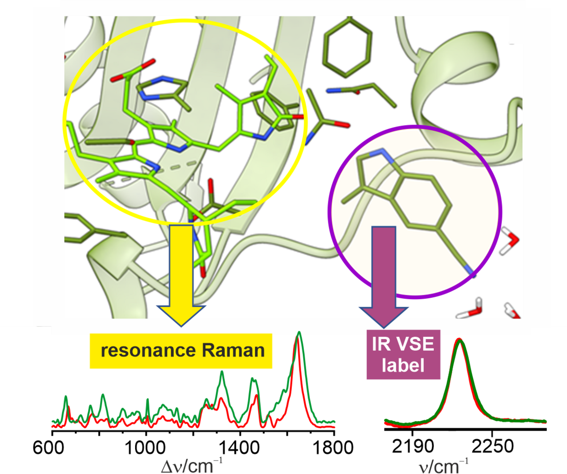
In this project, we studied representatives of both classes of photoreceptors by using a highly interdisciplinary approach involving various approaches from spectroscopy, molecular biology, and theory. As a sensory photoreceptor we have chosen a light-regulated enzyme from the green algae Synechocystis. This protein, Slr1393, carries a 'tetrapyrrole' chromophore derived from heme and switches between a red-light absorbing and a green-light absorbing form when it aborts light Here we were particularly interested on the role of individual amino acids for the structural and mechanistic properties of Slr1393. Accordingly, we replaced a critical tryptophan amino acid at position 496 with other natural amino acids and, as a particular challenge, non-canonical amino acids which were introduced via re-programming the genetic code. These synthetic amino acids carry a chemical group that can sense local electric fields, and thus serve as unique markers for infrared spectroscopy. This and other spectroscopic techniques were employed to selectively probe tryptophans and the tetrapyrrole chromophore upon switching between the red- and green-absorbing states. The results confirm x-ray crystallographic data that Trp496 interacts with the tetrapyrrole in the dark state, and separates from it in the illuminated state, but not by flipping into the solvent as had been suggested. In addition, Trp496 was found to play an important role for the reaction mechanism. In the second sub-project, we studied the membrane-bound bacterial rhodopsin from Krokinobacter eikastus (KR2). KR2 includes a retinal chromophore attached covalently to the protein.
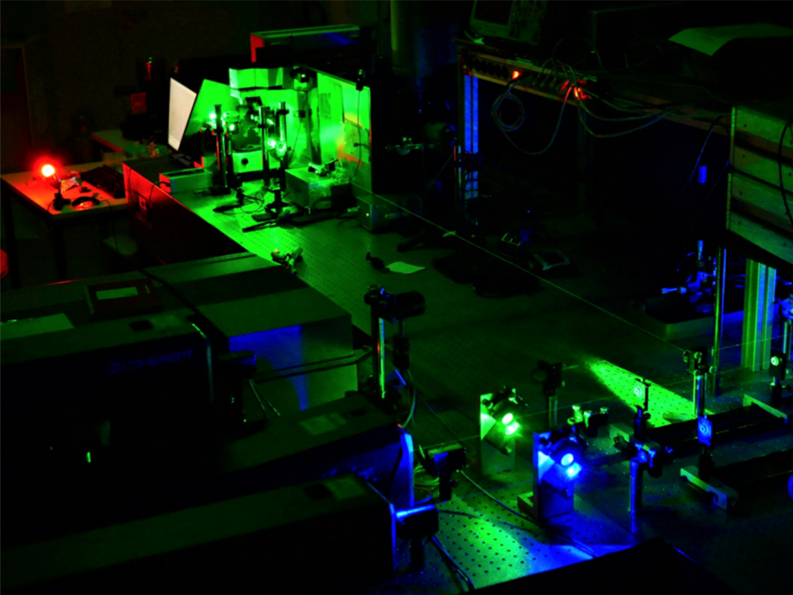
Upon light absorption, KR2 runs through a photocycle that is linked to the active transport of sodium across the membrane. The photocycle ends with the recovery of the parent state within ca. one second. Here we mainly employed time-resolved RR spectroscopy to monitor the temporal evolution of the various intermediate states during the photocycle. These studies not only aimed at deeper insight into structure-dynamics relationships of this photoreceptor, but also at a methodological development in time-resoved RR spectoscopy to overcome a series of drawbacks associated with previous approaches. Following optimization for KR2, the present set-up was employed for other retinal proteins, demonstrating its versatility for time-resolved RR experiments of photoreceptors.
Altogether, the present project provided important results for a deeper understanding of the tetrapyrrole protein Slr1393 and the retinal protein KR2. The findings are important also in a wider context of the research on photoreceptors in general and with respect to optimized analytical techniques.